

![]() canning a fluid-filled petri dish under the microscope, physician Stephen Boyers pauses when a small cluster of cells comes into view. "That's a nice two," he says, slowly adjusting the microscope's focus on the cluster. "That could be a three or four," he adds, checking a second cluster nearby.
canning a fluid-filled petri dish under the microscope, physician Stephen Boyers pauses when a small cluster of cells comes into view. "That's a nice two," he says, slowly adjusting the microscope's focus on the cluster. "That could be a three or four," he adds, checking a second cluster nearby.
 Each of the tiny clusters, just two, three or four cells in size, is the result of one of the most wondrous events in nature--fertilization of an egg by a sperm cell. Miraculously, these fertilization events,involving human cells, occurred in a small laboratory dish.
Boyers, who is chief of the Assisted Reproductive Technologies Program at the UC Davis Medical Center, is at a critical step in the technology of human in vitro fertilization: the selection of embryos for transfer to the uterus. Two days earlier, eggs were surgically removed from a woman's ovaries and placed in a carefully prepared broth called human tubal fluid. Six hours after that, sperm cells from her husband were added. The following day, evidence of fertilization of some of the eggs was detected, about 14 to 18 hours after introduction of the sperm cells.
Now, one day after fertilization, Boyers selects four embryos that are actively dividing and carefully inserts them into the woman's uterus through her cervix. In another eight to 10 days, a laboratory pregnancy test will tell her if an embryo has successfully attached to her uterine wall. Within the medical center's program, the average rate of achieving a pregnancy, regardless of the woman's age, is 35 percent.
That such a remarkable procedure as in vitro fertilization-embryo transfer can be performed and result in a term pregnancy speaks to the tremendous advances made in recent decades in our understanding of human reproductive biology. Among those contributing to the field are dozens of physicians and scientists working at clinics and laboratories throughout UC Davis. Many of them rely on nonhuman species to learn more about human reproduction--an area of study that is often difficult to approach because of ethical issues. Moving back and forth between animal models and clinical settings, they reveal fascinating clues to the mysteries of human procreation.
*
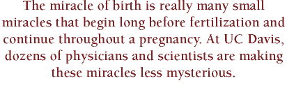
![]() or fertilization of a human egg to occur, whether it be in a petri dish or nature's setting--a woman's fallopian tube--a series of intricate events must occur to produce a viable egg in the woman and healthy sperm cells in the man. In women, development of a ripe egg and its release into a fallopian tube, a process called ovulation, is controlled by hormones produced by the brain, pituitary gland and ovaries.
or fertilization of a human egg to occur, whether it be in a petri dish or nature's setting--a woman's fallopian tube--a series of intricate events must occur to produce a viable egg in the woman and healthy sperm cells in the man. In women, development of a ripe egg and its release into a fallopian tube, a process called ovulation, is controlled by hormones produced by the brain, pituitary gland and ovaries.
According to Jeffrey Chang, professor of obstetrics and gynecology, the leading cause of abnormal ovulation is polycystic ovary syndrome. "The syndrome exists in about three to four percent of women of reproductive age," says Chang, who has studied the disorder for more than 20 years. "Those who have the syndrome usually experience infrequent or irregular menstrual periods, excessive hair growth and infertility."
 The subject of considerable research worldwide, polycystic ovary syndrome is now known to be a multisystem disease that involves the three parts of the body that control ovulation (the brain, specifically the hypothalamus; the pituitary gland; and the ovary) as well as other factors. At UC Davis, Chang and his colleagues have been studying the effects of reproductive steroid hormones and relevant protein hormones, such as insulin-like growth factor, on the ovary and pituitary gland in afflicted women. (Chang's interest in this factor stems from his earlier observation that patients with the syndrome have increased insulin levels.)
"No one really knows precisely why women with polycystic ovary syndrome fail to ovulate regularly," Chang says. "We do know that they exhibit abnormal gonadotropin [reproductive hormone] secretion patterns, which may be the key to failed ovulation. But to take the brain, pituitary, ovary and various proteins and put them into an understandable concept has been difficult."
By identifying the role each system and molecule plays in abnormal ovarian function, physicians such as Chang will be better able to develop treatment regimes for patients with the polycystic ovary syndrome. "It's difficult to treat infertility in these women if we don't understand what the ovary is doing," he notes.
*
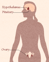
![]() hen ovulation does occur, the newly released egg enters a fallopian tube, ready to encounter sperm cells that have made it through the selective hurdles of a woman's reproductive tract. "What happens between the time sperm enter the woman and the time they meet the egg is critical," says James Overstreet, a professor in the Department of Obstetrics and Gynecology and director of the Institute of Toxicology and Environmental Health. "Reproductive technology has shown that you can 'cure' many kinds of infertility, including unexplained infertility, by putting eggs and sperm together in a dish, essentially bypassing the female reproductive tract."
hen ovulation does occur, the newly released egg enters a fallopian tube, ready to encounter sperm cells that have made it through the selective hurdles of a woman's reproductive tract. "What happens between the time sperm enter the woman and the time they meet the egg is critical," says James Overstreet, a professor in the Department of Obstetrics and Gynecology and director of the Institute of Toxicology and Environmental Health. "Reproductive technology has shown that you can 'cure' many kinds of infertility, including unexplained infertility, by putting eggs and sperm together in a dish, essentially bypassing the female reproductive tract."
 One hurdle that sperm cells encounter is the cervix. "We've shown that the cervix is a biological filter," Overstreet says. "Cervical mucus selects against sperm with abnormal shapes by presenting greater resistance to them than to normally shaped sperm." He notes that semen contains many abnormally shaped sperm cells, even in fertile men.
In some unprecedented studies, Overstreet and his research colleagues recovered sperm cells from cervical mucus and studied their physiology in the laboratory. They found that sperm cells have the same physiologic characteristics, such as motility, regardless of whether they have been in the cervix for one hour or 72 hours. Overstreet says that this finding supports the idea that the cervix can serve as a reservoir of sperm cells--a privileged environment that supports sperm survival.
Reproductive biologists suspect that more selection occurs further along a woman's reproductive tract, but there is far less data on other reproductive hurdles, and almost no data in humans. "We try to use a nonhuman primate model to address questions that we can't address in human research," explains Overstreet. "For example, for questions about the physiology of sperm in the uterus and oviduct [fallopian tube], we use cynomolgus monkeys as a model species."
Research such as Overstreet's has earned UC Davis a reputation for excellence in the study of human sperm. The campus's andrology laboratory was selected by the National Institutes of Health as the quality assurance site for a current multicenter study on the effectiveness of artificial insemination and superovulation as therapies for male infertility and unexplained infertility. The laboratory standardizes methods to assess sperm quality, trains technical staff and conducts quality assurance testing, all of which are essential in a multicenter study. UC Davis is also one of the six clinical research sites participating in the project.
*
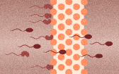
![]() nce a viable egg is produced and sperm cells have entered the fallopian tubes, fertilization is possible--if the two types of sex cells successfully meet. "Sperm have the opportunity to interact with many other kinds of cells in the female," says Jerry Hedrick, a biochemist in the Division of Biological Sciences. "How egg and sperm recognize one another is a fundamental question in reproductive biology."
nce a viable egg is produced and sperm cells have entered the fallopian tubes, fertilization is possible--if the two types of sex cells successfully meet. "Sperm have the opportunity to interact with many other kinds of cells in the female," says Jerry Hedrick, a biochemist in the Division of Biological Sciences. "How egg and sperm recognize one another is a fundamental question in reproductive biology."
Hedrick is trying to answer this question using approaches in biochemistry and molecular biology, along with nonhuman species to model the human system. "In biology, it is a dictum that you pick the simplest system that has the characteristics you want to study. Human reproduction is complex and female gametes [eggs] are hard to come by. We frequently turn to the frog as a model because the interaction of sperm and eggs in frogs appears to be similar to that in humans, and frog eggs are easy to obtain."
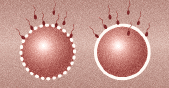 Hedrick's research has focused on four general recognition events that occur between sperm and egg: sperm binding to the egg surface; sperm acrosome reaction (changes in the sperm that enable it to fertilize an egg); sperm penetration of the egg; and the block to polyspermy, a process that prevents more than one sperm from entering and fertilizing the egg. Hedrick says that his laboratory has made the most progress on that last event. "We've discovered a new molecule in frogs that appears to function as one of two locks on the egg's door," he explains. "The block to poly-spermy is so important to the future existence of a new organism that nature appears to have created more than one mechanism to lock the door on other sperm."
Observations such as this lead Hedrick to wonder whether there are multiple mechanisms to block polyspermy in mammals. He is currently moving up the evolutionary tree, looking for a molecule in mammals that is comparable in function to the one his laboratory discovered in frogs. The discovery of a similar molecule in humans could present reproductive biologists with an opportunity to develop a new contraceptive. By creating a molecular "magic bullet" that binds specifically to the molecule, scientists could possibly disrupt the normal events of fertilization.
*
![]() ontrary to an impression held by many, a fertilized egg does not simply float down a woman's fallopian tube en route to her uterus. Rather, the egg is guided along its narrow path by the beating movement of hairlike structures called cilia that project from cells lining the fallopian tubes. And, when the egg arrives at the uterus, it enters a moist, flat inner space--not a fluid-filled cavity.
ontrary to an impression held by many, a fertilized egg does not simply float down a woman's fallopian tube en route to her uterus. Rather, the egg is guided along its narrow path by the beating movement of hairlike structures called cilia that project from cells lining the fallopian tubes. And, when the egg arrives at the uterus, it enters a moist, flat inner space--not a fluid-filled cavity.
By the time a fertilized egg reaches the uterus, usually a few days after conception, it has already divided many times and has become organized into a hollow sphere of cells technically called a blastocyst, but also known as an embryo. It is the blastocyst that implants into the inner surface of the uterus, the endometrium, and initiates the development of a placenta.
During the period of embryonic development that begins with fertilization and ends with successful implantation of the blastocyst--known as "preimplantation development"--up to 50 percent of human conceptions fail to survive, says Lynn Wiley, professor of obstetrics and gynecology. One reason for this high failure rate is the inability of an embryo to implant. "Only certain cells within the embryo can implant and form a placenta. Without these cells, or if these cells are not healthy, implantation will fail," she says.
Wiley's research laboratory uses the mouse as a model to study developmental changes that occur during preimplantation and to investigate environmental factors that cause embryo failure before implantation. Some likely factors are dioxins, which are by-products of combustion processes used in manufacturing. "Dioxins happen to be especially toxic to the developing placenta," Wiley says. "Mouse embryos exposed to dioxins suffer around a 70 percent implantation loss. Dioxins also appear to be more toxic to male embryos than to female embryos."
Reproductive biologists like Wiley have found the mouse to be a useful model for the study of purported reproductive toxicants. The early mouse embryo is about the same size as the human embryo; it can be cultured from fertilization to implantation stages, just as human embryos are in in vitro fertilization–embryo transfer procedures; and it appears to obey the same rules in developing the capacity to form a placenta. However, there are some differences, notes Wiley. "The human embryo can implant anywhere inside the body cavity [producing an ectopic pregnancy], while the mouse embryo is more discriminating about where it can implant. Human embryos are also much less picky about the timing of their arrival in the uterus and their stage of embryonic development. Mouse embryos follow much stricter rules in this regard."
*
![]() llen Enders, professor emeritus of cell biology and human anatomy in the School of Medicine, has focused much of his research on the cellular events that occur during implantation and the early formation of a placenta. Yet, despite his work and that of others in the field, these events are still not well-understood, particularly in humans.
llen Enders, professor emeritus of cell biology and human anatomy in the School of Medicine, has focused much of his research on the cellular events that occur during implantation and the early formation of a placenta. Yet, despite his work and that of others in the field, these events are still not well-understood, particularly in humans.
"It is extremely difficult to get material that is early enough to study the process of implantation and placenta formation," explains Enders. "Most of what we know comes from a stained slide collection, the Carnegie collection, prepared in the 1940s and 1950s." The collection was maintained at UC Davis for several years, and during that time Enders studied and photographed the slides extensively. "The earliest stage in the collection is after the blastocyst has penetrated the epithelium [the layer of cells that lines the endometrium]. Nobody has ever seen in the human the first attachment of the blastocyst to the uterus."
Because it is so difficult to obtain human tissue, Enders turns to animal models to investigate why and how the blastocyst "sticks" to the uterus. He finds animal models particularly useful if the animal shares a reproductive characteristic with humans that is easy to study in the animal. For example, one model, the spotted skunk, has a relatively large blastocyst and several of them, so it is easy to identify and study implantation sites. However, he says that caution should be used when drawing conclusions from comparative animal studies. "Things are not exactly the same, and while some differences may not be significant, some may be."
*
![]() he first clinical sign of implantation and pregnancy in humans is a missed menstrual period. "A woman gets her first hint that she is pregnant about two weeks after conception," says Steven Nakajima, assistant professor of obstetrics and gynecology. "Because we don't know exactly when a woman gets pregnant, it's been difficult to study the events of very early pregnancy."
he first clinical sign of implantation and pregnancy in humans is a missed menstrual period. "A woman gets her first hint that she is pregnant about two weeks after conception," says Steven Nakajima, assistant professor of obstetrics and gynecology. "Because we don't know exactly when a woman gets pregnant, it's been difficult to study the events of very early pregnancy."
Nakajima and his colleagues are characterizing hormonal activity associated with early pregnancy in a unique prospective study supported by the Superfund, a federal program established in 1980 to clean up the United States' most seriously polluted toxic waste sites. (A small part of the program funds basic research on human health.) The study participants are women who are trying to conceive through donor insemination. Each woman donates a blood and urine sample every day from the day of insemination to the day she begins her menstrual period. If a woman misses her period, samples are taken for another 10 to 12 days. In addition, a series of blood samples is taken eight days after the woman ovulates, when implantation is likely to occur. By measuring the amount of selected hormones in each sample, Nakajima is developing a profile of normal hormonal activity during implantation and early pregnancy. "What's inherently exciting about this is that we are trying to characterize an event that has been with us since the dawn of time, and yet we don't know very much about it," he says.
With baseline information about normal conception in hand, Nakajima has been able to turn his attention to studies on abnormal processes such as repetitive miscarriage. "We're using our data from the normal pregnancy state to mimic pregnancy in women who have had miscarriages," he explains. Each study participant wears for three days a battery-operated pump that infuses the pregnancy hormone, human chorionic gonadotropin [hCG], into a vein in a pattern that mimics a normal pregnancy. "We make them pseudopregnant and see whether or not their ovary responds the way it should in a normal pregnancy. If we identify someone who doesn't respond normally to the hCG stimulus, we can treat her prospectively and give her a little bit more hope."
*
![]() he events of early reproduction are among the most complex and least understood in human biology. They are also some of the most fascinating and challenging areas of contemporary research, engaging scientists and clinicians from a wide variety of disciplines. Like many reproductive biologists, the investigators identified here are driven to explore the processes that enable two types of cells, sperm cell and egg, to join, develop into an embryo and implant in the womb. They also share with many of us a fundamental desire to know and understand the events that ensure the continuation of human life.
he events of early reproduction are among the most complex and least understood in human biology. They are also some of the most fascinating and challenging areas of contemporary research, engaging scientists and clinicians from a wide variety of disciplines. Like many reproductive biologists, the investigators identified here are driven to explore the processes that enable two types of cells, sperm cell and egg, to join, develop into an embryo and implant in the womb. They also share with many of us a fundamental desire to know and understand the events that ensure the continuation of human life.
*
Karen Guin is a public information officer in the UC Davis Division of Biological Sciences.